Super Stiff PTFE Guidewire Percutaneous Nephrostomy Set with Radiopaque Markers 08-22Fr
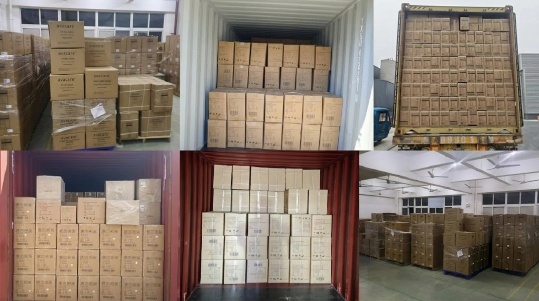
Order MOQ: 50000 pcs
Size :08-22Fr
Private Label: OEM and ODM
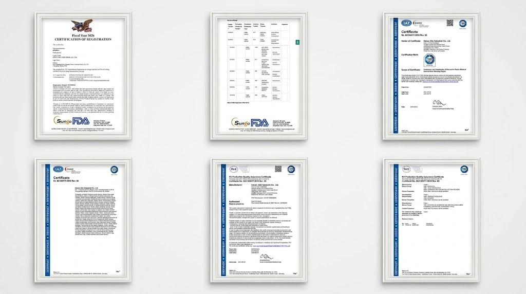
Quality Certifications: CE / ISO13485/ USA FDA
Delivery Time: 30–45 Days (approx.)
Payment Methods: T/T or L/C Accepted
Super Stiff PTFE Guidewire Percutaneous Nephrostomy Set with Radiopaque Markers 08-22Fr
HENAN AILE INDUSTRIAL CO., LTD with over 12 years of medical experience.
We hold certifications, including CE, ISO 13485 & USA FDA.
1. Key Advantages &Product Overview Key Advantages:
High Columnar Track Stability (Super Stiff Core): The high-tensile core wire prevents bucking, kink deformation, and guidewire whipping when advancing stiff dilators across dense lumbodorsal fascia, securing access inside the target calyx.
Low-Friction Thermal PTFE Coating: The dry fluoropolymer PTFE jacket dramatically cuts kinetic and static surface friction, ensuring effortless dilator travel without binding or hydraulic stiction.
Precise Multi-Modal Radiopacity: Featuring precision-banded radiopaque markers along the distal guidewire segment and drainage catheter loop, clinicians achieve real-time positional verification under fluoroscopy and echogenic acoustic shadowing under ultrasound.
Graduated Coaxial Dilatation (8–22 Fr): The fascial dilators transition smoothly from 8 Fr to 22 Fr with seamless atraumatic tip bevels, minimizing dilator-guidewire gaps and preventing shear stress on the renal parenchyma.
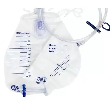
Turnkey Sterile Procedural Integration: Combines puncture needles, guide tracks, sequential dilators, retention catheters, securement devices, and fluid adapters within an integrated sterile tray, optimizing operating room turnaround.
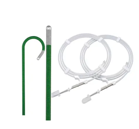
The Super Stiff PTFE Guidewire Percutaneous Nephrostomy Set with Radiopaque Markers 08-22Fr is an all-in-one interventional urology kit engineered for secure percutaneous renal access, rapid pelvic decompression, and seamless working channel creation.
Designed to execute the standardized Seldinger technique with clinical confidence, the set incorporates a heavy-duty Super Stiff PTFE-Coated Guidewire, a graduated series of atraumatic fascial dilators spanning 8 Fr to 22 Fr, high-clarity radiopaque positioning markers, and a locking nephrostomy drainage catheter. This cohesive system delivers maximum track stability, mitigates tissue trauma across muscular and fascial barriers, and provides superior anatomical control during minimally invasive procedures like Percutaneous Nephrolithotomy (PCNL).

2. Product Specifications Table
| Parameter / Feature | Detailed Specifications |
| Product Model | Super Stiff PTFE Guidewire Percutaneous Nephrostomy Set with Radiopaque Markers 08-22Fr |
| Dilator Size Coverage | 8 Fr to 22 Fr in progressive increments (8, 10, 12, 14, 16, 18, 20, 22 Fr) |
| Guidewire Dimensions | Outer Diameter: 0.038" (0.97 mm); Lengths: 80 cm / 100 cm / 150 cm |
| Core & Surface Coating | High-elasticity spring stainless steel/nitinol core; baked medical-grade PTFE resin |
| Distal Guidewire Tip | Flexible atraumatic safety tip (Straight or 3 mm / 6 mm J-Tip options) |
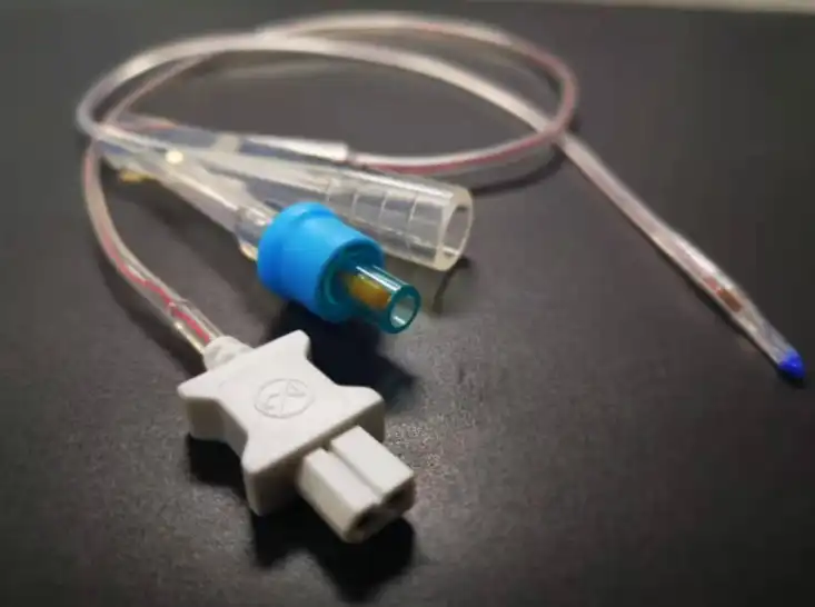
| Catheter Configurations | Locking Pigtail Catheter with tether string / Malecot (four-wing retention) |
| Imaging Markers | Distal radiopaque guidewire band + full-length barium-impregnated stripe & rings |
| Access Needle | 18G two-piece/three-piece echogenic-tip trocar/Chiba needle (20 cm working length) |
| Component Polymers | Medical Thermoplastic Polyurethane (TPU) / High-Density Polyethylene (HDPE) |
| Sterilization & Pack | 100% Ethylene Oxide (EO) gas sterilized; sterile medical Tyvek peel blister pack |
| Quality Compliance | ISO 13485 manufacturing standards, CE MDR cleared, FDA registered |
3. Material Comparative Analysis: Engineering Properties
Performance in interventional endourology depends heavily on substrate selection across guidewires, dilators, and indwelling lines:
-
Guidewire Core Architecture: Super Stiff PTFE vs. Hydrophilic Nitinol (Zebra):
-
Super Stiff PTFE Guidewire (This Set): Delivers superior axial rigidity and columnar support. It resists lateral buckling under push-forces when traversing dense muscular planes, keeping the access track stable during dilation. The sintered PTFE coating remains firmly adhered without peeling.
-
Hydrophilic Zebra Guidewire: Features high lubricity when wet, making it suitable for negotiating tortuous ureteral strictures. However, its lower radial stiffness allows bowing during percutaneous tract dilation, risking accidental guidewire displacement or false-passage dissection.
-
-
Polymer Tubing: Medical TPU vs. HDPE vs. Conventional PVC:
-
Medical Polyurethane (TPU Catheter): Features thermo-responsive softening behavior. The catheter maintains structural stiffness during insertion, then softens at body temperature inside the renal pelvis to reduce localized tissue pressure. It resists encrustation and is optimized for intermediate dwell times.
-
High-Density Polyethylene (HDPE Dilators): Offers high structural integrity and low wall friction, making it the ideal material for sequential dilators navigating fibrotic scar tissue.
-
Standard PVC: Less flexible over time and reliant on chemical plasticizers, standard PVC is prone to embrittlement and encrustation during renal urinary stasis.
-

4. Clinical Applications & Target Patient Demographics
Core Clinical Indications:
-
Percutaneous Nephrolithotomy (PCNL / Mini-PCNL): Creating and expanding a stable working tract from the flank skin into the renal collecting system for endoscopic nephroscope insertion and lithotripsy.
-
Emergency Pyelocaliceal Decompression: Diverting infected urine and relieving pressure during acute hydronephrosis secondary to ureteral calculi, retroperitoneal fibrosis, or pelvic tumors.
-
Diagnostic & Therapeutic Interventions: Antegrade pyelography, antegrade double-J stent insertion, endopyelotomy, and balloon dilatation of ureteropelvic junction (UPJ) obstructions.
Target Demographics:
-
Patients with post-renal obstructive uropathy presenting with rising serum creatinine or sepsis.
-
Urolithiasis candidates requiring percutaneous surgical stone extraction.
-
Individuals where retrograde transurethral catheterization has failed due to altered anatomy, ureteral strictures, or prostatic hypertrophy.
5. Why Choose Our Super Stiff PCN Access Set?
-
Zero-Buckle Track Assurance: The high-modulus core wire eliminates guidewire looping in the retroperitoneal space, ensuring smooth, predictable dilation along the primary access path.
-
Atraumatic Sequential Dilation: Precision-beveled dilator tapers eliminate step-transitions against the 0.038" wire, lowering entry friction and preventing caliceal perforation.
-
Continuous Radiographic Traceability: High-contrast barium lines and distal radiopaque markers keep the tip position visible under fluoroscopic surveillance, preventing unintended over-advancement.
-
Complete Procedural Versatility: The 8–22 Fr kit covers both small-caliber acute drainage protocols and broad-caliber endoscopic tract dilatation.
6. (FAQ)
Q1: Why is a "Super Stiff" guidewire required instead of a standard wire in PCN procedures?
A1: Penetrating through the skin, lumbar fascia, and renal parenchyma requires significant axial force. A standard wire bends under this resistance, which can shift the trajectory or dislodge the wire from the calyx. A super-stiff wire provides the rigid rail needed to keep dilators centered.
Q2: Will the PTFE coating flake or peel off during tough fascial dilations?
A2: No. The PTFE coating is thermally sintered to the core wire. As long as the wire does not rub directly against the sharp cutting edge of an access needle or scalpel, normal dilator travel will not cause delamination.
Q3: Can the radiopaque markers be visualized under standard ultrasound?
A3: Yes. In addition to clear radiopacity on fluoroscopy screens, the structural density of the marker bands and the needle's textured tip generate distinct hyperechoic acoustic reflections with acoustic shadowing under ultrasound guidance.
Q4: How should clinicians determine the final dilator size between 8 Fr and 22 Fr?
A4: An 8 Fr to 12 Fr channel is typically sufficient for temporary decompression or purulent drainage via a pigtail catheter. If the goal is PCNL stone management, dilation is advanced up to 16 Fr–22 Fr to place an appropriately sized working sheath.
Q5: What is the correct protocol for safely removing a locking pigtail catheter?
A5: The locking retention suture must be snipped at the outer connector hub to release tension on the internal thread. Confirm the distal pigtail loop has straightened out before extracting the tube along the tract to avoid mucosal tearing.
Q6: What imaging modality is recommended for initial 18G puncture needle placement?
A6: The 18G needle features depth graduations and an echogenic-enhanced tip suited for direct real-time ultrasound guidance, single-plane fluoroscopy, or combined bi-modal navigation.
Q7: What steps should be taken if unexpected resistance occurs during sequential dilation?
A7: Stop advancing the dilator immediately and check fluoroscopy to verify guidewire trajectory and ensure it has not kinked within the calyx. If the restriction is caused by dense superficial fascia, use the scalpel to extend the skin incision by 1–2 mm adjacent to the wire.
Q8: What is the safe indwelling lifespan of the included nephrostomy drainage tube?
A8: The medical-grade TPU catheter is indicated for indwelling durations up to 30 to 90 days, depending on patient encrustation risk and urine sediment. For long-term diversion, scheduled over-the-wire tube exchanges are recommended.



_1782811410038.webp)